Từ Blaivas M, Theodoro D, Sierzenski PR: Elevated intracranial pressure detected by bedside emergency ultrasonography of the optic nerve sheath. Acad Emerg Med. 2003 Apr;10(4):376-81.
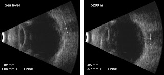
Đo đường kính dây thần kinh thị giác sau nhãn cầu bằng siêu âm là cách thức đơn giản, không xâm lấn và có hiệu quả trong phát hiện tăng áp lực nội sọ. Blaivas và cs (2003) đã dùng siêu âm nhãn cầu để đánh giá tăng áp lực nội sọ ở người lớn tại khoa cấp cứu. Bệnh nhân mất tri giác nhiều mức độ có thể bị tăng áp lực nội sọ do nhiều nguyên nhân. Tăng áp lực nội sọ ở khoa cấp cứu thường xảy ra ở bệnh nhân chấn thương đầu hoặc bị xuất huyết nội sọ tự phát.
Phù gai thị thường xảy ra muộn nhiều giờ sau tăng áp nội sọ. Có một phương tiện khám nhanh chóng, tại giường và không xâm lấn là yêu cầu cần thiết để phát hiện tăng áp nội sọ khi không có phương tiện chẩn đoán hình ảnh quy ước hữu hiệu.
Dây thần kinh thị giác ở sau nhãn cầu và được bọc bằng một bao chứa dịch, bao này tiếp giáp với màng cứng và khoang dưới nhện trong có dịch não tủy lưu thông. Tương quan giữa đường kính bao dây thần kinh thị giác và áp lực nội sọ đã được công nhận. Đo đường kính dây thần kinh thị giác có thể phát hiện tăng áp lực nội sọ.
Trên siêu âm, với đầu dò linear 7,5-10MHz, đường kính dây thần kinh thị giác bình thường khoảng 5mm. Bao dây thần kinh thị giác được đo sau nhãn cầu 3mm cả 2 bên mắt. Yêu cầu của vị trí 3mm sau nhãn cầu do tại đây sự tương phản của siêu âm là rõ nhất, và kết quả có thể lập lại. Trung bình nên đo 2 kích thước. Khi đường kính bao dây thần kinh thị giác lớn hơn 5mm có thể xem như bất thường và chẩn đoán tăng áp lực nội sọ có thể đặt ra.
Bao dây thần kinh thị giác giãn 5,3mm ở bệnh nhân tăng áp nội sọ. Đo khoảng cách sau nhãn cầu 3mm và kích thước đo lần sau là đường kính bao dây thần kinh thị giác = 5,3mm (Hình của Michael Blaivas, M.D.)
Báo cáo của Blaivas và cs phát hiện 14/35 bệnh nhân có tăng áp lực nội sọ bằng siêu âm và xác chẩn bằng CT. Đường kính bao dây thần kinh thị giác trong tăng áp lực nội sọ là 6,27mm (95% CI =5,6-6,89). Số còn lại, không có tăng áp lực nội sọ, là 4,42 mm (95% CI = 4,15 to 4,72). Độ nhạy và độ đặc hiệu lần lượt là 100% và 95%, giá trị tiên đoán dương và âm tính là 93 và 100%.
ASC20110868
Title Ultrasound Measurement Of Optic Nerve Sheath Diameter Estimation Of Intracranial Pressure In Adult Trauma Patients
L. Yeung- R. Kwan- A. Strumwasser-- E. Cureton- K. Dozier- E. Miraflor- J. Sadjadi- G. Victorino
UCSF-East Bay Department Of Surgery - Alameda County Medical Center- 1411 E. 31st Street #OA2 -Oakland, CA USA
Abstract
Introduction: Ultrasound measurement of optic nerve sheath diameter (ONSD) can be used as a rapid non-invasive assessment tool for evaluating intracranial pressure (ICP). We hypothesized that ultrasound measurement of ONSD accurately predicts ICP before and after therapeutic efforts to control ICP are performed. The aims of this study were
1) to correlate ultrasound measurement of ONSD to subsequent invasive ICP measurement, and
2) to correlate the side of head injury with ipsilateral and contralateral ONSD and invasive ICP measurement.
Methods: A blinded, prospective, clinical study of adult trauma patients with blunt or penetrating head trauma requiring an intracranial pressure monitor was performed at a University-based urban trauma center. Left and right optic nerve sheath diameters were measured by optic ultrasound before and after invasive placement of an ICP monitor (Camino bolt or ventriculostomy).
Results: 54 independent evaluations of ONSD in 9 trauma patients (8 blunt, 1 penetrating) requiring an intracranial pressure monitor for head trauma were enrolled. Pre-ONSD diameter (3 recordings per patient = 27 assessments) and post-ONSD diameter (3 recordings per patient = 27 assessments) were assessed and correlated with side of injury in the presence of an ICP monitor. There was no difference between left (mean pre-ONSD = 0.64 ± 0.07cm vs. mean post-ONSD = 0.60 ± 0.04cm, p=0.7), right (mean pre-ONSD = 0.62 ± 0.07cm vs. mean post-ONSD = 0.60 ± 0.04cm, p= 0.8), pre- and post-intervention ONSD measurements. No correlation was observed between invasive ICP measurements and pre-intervention ONSD (pre-intervention left vs. ICP R2 = 0.42, pre-intervention right vs. ICP R2 = 0.43), regardless of the side of traumatic head injury, pressure monitor location, or presence or absence of elevated intracranial pressure (P over 0.11 for each). Moreover, there was no correlation observed between invasive ICP measurements and post-intervention ONSD (post-intervention left vs. ICP R2 = 0.52, post-intervention right vs. ICP R2 = 0.52), regardless of the side of traumatic head injury, pressure monitor location, or presence or absence of elevated intracranial pressure (P over 0.15 for each). Ultrasound ONSD had a sensitivity of 50% and an accuracy of 37.5% for predicting elevated ICP.
Conclusions: Despite the expanding use of ultrasound in evaluating trauma patients, the clinical utility of ocular ultrasound measurement of optic nerve sheath diameter to predict ICP in head injured patients prior to invasive monitoring is not a reliable test due to variability and lack of correlation.


Không có nhận xét nào :
Đăng nhận xét