Abstract
Although elastography can enhance the differential diagnosis of thyroid nodules, its diagnostic
performance is not ideal at present. Further improvements in the technique and
creation of robust diagnostic criteria are necessary. The purpose of this study
was to compare the usefulness of strain elastography and a new generation of
elasticity imaging called supersonic shear wave elastography (SSWE) in differential evaluation of thyroid nodules. Six thyroid nodules in
4 patients were studied. SSWE yielded 1 true-positive and 5 true-negative
results. Strain elastography yielded 5 false-positive results and 1
false-negative result. A novel finding appreciated with SSWE, were punctate foci of increased stiffness corresponding to microcalcifications in 4 nodules, some not
visible on B-mode ultrasound, as opposed to soft, colloid-inspissated areas
visible on B-mode ultrasound in 2 nodules. This preliminary paper indicates
that SSWE may outperform strain elastography in differentiation
of thyroid nodules with regard to their stiffness.
SSWE showed the possibility of differentiation of high echogenic foci into
microcalcifications and inspissated colloid, adding a new dimension to
thyroid elastography. Further multicenter large-scale studies of thyroid
nodules evaluating different elastographic methods are
warranted.
Methods
During a few weeks trial time in 2010, four consecutive
patients with single thyroid nodule (n = 1) and nodular goiter (n = 3) were
evaluated. Approval for this study was obtained from the Ethics Committee of
the Medical University of Warsaw
The Bmode and power Doppler ultrasound of whole thyroid and
neck lymph nodes was performed. Six dominant thyroid nodules (in regard to
B-mode and power Doppler ultrasound features) were evaluated with shear wave
and strain elastography qualitatively and quantitatively as well as some with contrast-enhanced ultrasound (Sonovue (Bracco)). The
examinations were performed with following scanner: AiXplorer (Supersonic Imagine
Inc. France)—SSWE, Aplio XG (Toshiba , Japan )—strain elastography, Technos (Esaote , Italy
The final diagnosis was based on clinical evaluation,
multiple FNB, 1 year followup, or surgery.
Supersonic shear weave elastography consists of the
generation of remote radiation force by focused ultrasonic beams, the so-called
“pushing beams,” a patented technology called “Sonic Touch”. Using Sonic
Touch, ultrasound beams are successively focused at different depth in tissues. The source is moved at a speed that is higher than the speed of
the shear waves that are generated. In this supersonic regime, shear waves are
coherently summed in a “Mach cone” shape, which increases their amplitude and
improves their propagation distance. For a fixed acoustic power at a given location, Sonic Touch increases shear wave generation efficiency by a factor of 4 to 8
compared to a nonsupersonic source. After generation of this shear wave,
an ultrafast echographic imaging sequence is performed to acquire successive
raw radiofrequency dots at a very high-frame rate (up to 20,000 frames per
second). Based on Young’s modulus formula, the assessment of tissue elasticity
can be derived from shear wave propagation speed. A color-coded image is
displayed, which shows softer tissue in blue and stiffer tissue
in red. Quantitative information is delivered; elasticity is expressed in kilo-Pascal (kPa).
This preliminary paper based on small number of cases
indicates that SSWE indicated correctly thyroid nodules suspicious for cancer
in contrast to strain elastography. False positives on strain elastography
could be due to liquid or degenerative content of nodules.
However, imaging with SSWE, as a sensitive method of
evaluation of stiffness of human tissue, the operator
should be aware of physiological processes influencing the elasticity and easily
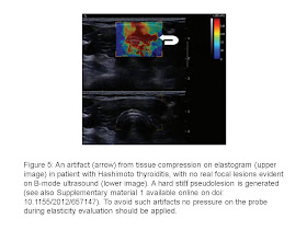
apply a few rules to avoid artifacts (Figures 4, 5 and 6). Among well-known artifacts on SSWE that should be
mentioned is the one that can be encountered in any region when the SSWE can be
applied: the increased stiffness of the structures under
externally applied pressure (Figures 4 and 5) that can be due to nonlinear elastic effects,
well explained by theory.
Another artifact that can be encountered in thyroid SSWE is
one of increased stiffness in the isthmus of the thyroid due
to trachea (Figure 6). It can be avoided with imaging in paracoronal plane of
the nodule that does not incorporate the trachea. However, it is important to
state that these artifacts when properly interpreted do not hinder the
accurate diagnosis.
Supersonic shear wave elastography may add a new dimension
to ultrasound evaluation of thyroid nodules in several ways, for example:
(a) improve general performance in elasticity differentiation of thyroid nodules over strain elastography due to
its high reproducibility, independence of examiners skill and numeral scale of
elasticity measurement in kPa;
(b) overcome the limitations of strain elastography=
(i) nodules with liquid components or with degenerative
changes;
(ii) small nodules (very good spatial resolution of the
technique);
(iii) large nodules (possibility of subsequent determination
of stiff regions even of large nodules,
without the need of visualizing the whole nodule on one image);
(iv) multinodular goiter with no or scarce normal thyroid
tissue as a reference;
(c) differentiation between soft-inspissated
colloid and stiff microcalcifications;
(d) visualization of microcalcifications, even not visualized
on B-mode imaging (may increase sensitivity and decrease specificity of thyroid
cancer diagnosis);
(e) introduction of three-dimensional elastographic images
to routine clinical practice and to national thyroid cancer databases, as
this technique is
already available and enables rapid acquisition of 3D
ultrasound and elastographic data. This would devoid diagnostic process and
data archiving of image selection bias attributable to 2D ultrasound
examination.
Further multicenter large scale studies of thyroid nodules
evaluating different elastographic methods are warranted,
including (a) investigation of developmental models of diseases that link
biomechanical properties (elastography findings) with genetic, cellular,
biochemical, and gross pathological changes; (b) comparison of accuracy of different elastographic methods; (c) establishment of optimal
diagnostic elastographic criteria; (d) establishment of limitations of
different elastographic methods in relation to evaluation of thyroid pathology.


Không có nhận xét nào:
Đăng nhận xét