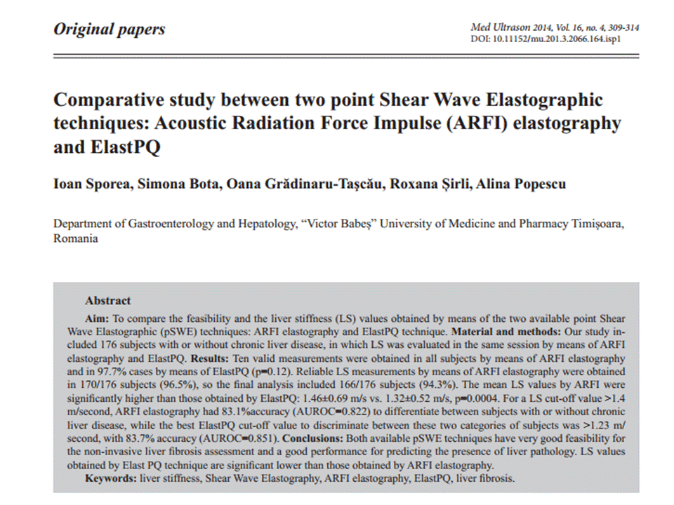
Liver stiffness values obtained by Elast PQ technique are
significantly lower than those obtained by ARFI elastography and future studies
are needed to establish the best LS cut-offs assessed by ElastPQ for predicting
different liver fibrosis stages.
--------------------------------------------------------------------------------------------------
ElastPQ
(Philips)
from JSUM
Ultrasound Elastography Practice Guideline Liver
Masatoshi Kudo1*,
Tsuyoshi Shiina2, Fuminori Moriyasu3, Hiroko Iijima4, Ryosuke Tateishi5,
Norihisa Yada1, Kenji Fujimoto6, 7, Hiroyasu Morikawa8, Masashi Hirooka9,
Yasukiyo Sumino10, Takashi Kumada11
A) Introduction
Name: ElastPQ (PQ:
point quantification)
Equipment: The iU22
xMATRIX ultrasound system (iU: Intelligent Ultrasound)
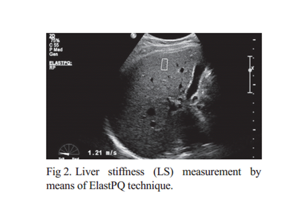
ElastPQ is a
non-invasive diagnostic tool to measure tissue stiffness using an ARFI-based
technology. Immediately after image acquisition, the screen displays the image
and measurement results, including the mean and median values and the
deviations in kPa or m/s (Fig. 33).
*If measurement reliability is low, 0.00 kPa will be
displayed as the result.
Elastic value E [kPa] is calculated using the equation
E = 3ρVsଶ where Vs
[m/s] is defined as the shear wave propagation velocity and ρ as tissue density
(whose approximated value in the human body is 1).
An ROI can be placed
anywhere but at a depth of uder 8 cm.
B) Indication
1. Quantitative
assessment of liver fibrosis in diffuse liver diseases
2. Neoplastic lesions of the liver
C) Procedures (including Tips and Tricks)
1. Perform right intercostal scanning to visualize the liver
2. Steadily place the
probe with minimum compression
3. Set the ROI with a
depth of under 8 cm.
4. Ask the patient to breath hold (if not possible, ask the patient to breathe as shallowly as possible).
5. Push the” Update” button for quantification
5. Push the” Update” button for quantification
6. The use of a mean value from more than 10 measurements is recommended.
Approach the right hepatic lobe from the right intercostal space. Avoid the left hepatic lobe because the measurement is affected by cardiac movement. Breath hold without exerting abdominal pressure. The most appropriate ROI is the center of the image, namely, immediately below the probe, and 3–5 cm from the probe surface. Avoid blood vessels, any necrotic areas, the boundary between organs, and areas influenced by cardiac movement (ex. left hepatic lobe). Three frequencies (R1/RP/P1) are available. The measurement sensitivity of areas deep inside the body can be improved by using a lower frequency.
D) Results (What does the value mean?)
・Healthy liver: 4 kPa (2.5–4.7 kPa, 1–1.5 m/s)
・Mild fibrosis: 7 kPa (4.7–12.0 kPa, 1.5–2.0 m/s)
・Moderate–severe fibrosis: 12 kPa (12.0–21.0 kPa, 2.0–2.5 m/s)
・Severe fibrosis: over 21
kPa (over 2.5 m/s)
E) Limitations
・There is a limit to
measurable depth.
・ElastPQ is affected by respiratory and body movement.
・Cardiac movement also affects the system.
・Accuracy of measurement depends on the skills of the
examiner.
・Measurement accuracy is generally low at the sides of an
image.
・Ribs may cast lateral acoustic shadows.
F) Recommendations
At present, the number of studies using ElastPQ is not large
enough to reach a definitive conclusion. We look forward to having more study
results in the near future.


Không có nhận xét nào :
Đăng nhận xét