Urinary bladder has many inherent characteristics that make it an ideal
structure for evaluating with three-dimensional (3D) volume ultrasound (US).
The purpose of this study is to evaluate the application of 3D sonography in
assessing bladder pathologies.
Materials and methods
One hundred patients were evaluated in this study. The cases were taken
from the pool referred for the evaluation of the renal system (kidney, ureter,
and bladder) abbreviated as US KUB at our hospital. Few selected observations
are presented here. The examination was performed with the bladder filled up to
250-350 ml, or wherever adequate distension was noted with wide separation of
the bladder walls. Routine (two-dimensional) 2D scanning was followed with the
acquisition of 3D volume using abdominal and endocavitary probes: RAB2-5L, 3D
abdominal (2-5 MHz), RIC5-9H endocavitary (5-9 MHz) using an US system. The
maximum field of view (FOV) was chosen to encompass the entire bladder volume
and to provide depth perception. High-resolution near-field images were
acquired using a smaller FOV. After collecting the 3D data, a surface rendering
algorithm was used for postprocessing to obtain cystoscopy-like US images of
the urinary bladder.
Conclusion
3D virtual cystoscopy is a promising technique for evaluating bladder
pathologies. Its multiplanar capabilities and surface rendering capabilities
are helpful for further characterizing the lesions seen on 2D US. It can serve
as a good road map prior to cystoscopy.
----
Results
The trigone region of the bladder and the distal ureters can
be seen in detail on the 3D-rendered images shown in Fig. 1 in all of our
evaluated patients.
An elderly gentleman who had complaints of increased frequency
of micturition was examined in this study.
Routine 2D US revealed an enlarged prostate gland. Using 3D
US, the indentation of the median lobe into the base of the bladder was clearly
observed (Fig. 2).
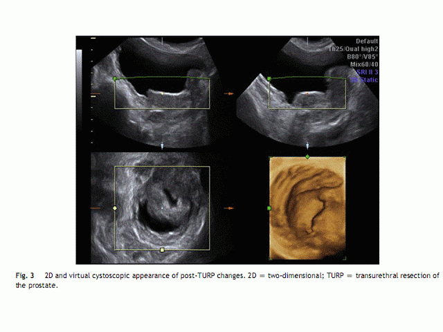
In another patient who had undergone transurethral resection
of the prostate (TURP), post-TURP changes were noted, similar to those in
cystoscopic findings (Fig. 3).
In this study, an ectopic ureteric opening with refluxive “golf-hole”
type appearance was localized (Fig. 4A). The patient was a 2-year-old girl
having recurrent urinary tract infections and hydronephrosis and hydroureter.
The micturating cystourethrogram showed Grade 3 reflux
(Fig. 4B).
In one patient who presented with hematuria, bladder mass
was observed. On using 3D US, one could better understand its morphology and
spatial orientation. This agreed well with the cystoscopic findings, as shown in
Fig. 5.
3D US also delineated diffuse bladder wall urothelial irregularity
in patients who experienced burning and urgency of micturition. The diagnosis
of cystitis was made, which correlated with urine microscopy, as shown in Fig.
6.
In one elderly patient with obstructive uropathy, diverticula
were observed in the 3D cystoscopic view, as shown in Fig. 7.
Using the inversion mode and surface rendering technique,
the bladder cast for morphological evaluation of the shape was obtained. One
interesting observation was the classical “Christmas tree” appearance of the
bladder in a child with a neurogenic bladder, as shown in Fig. 8A. This was an
8-year-old female with sacral agenesis (Fig. 8B).
In a 40-year-old female patient, using endovaginal volume
probe, one could observe the adder-head-type morphology of a ureterocele (Fig.
9). This patient’s earlier US had shown mild hydronephrosis on the affected side.
Discussion
Recent advances in computer technology and display techniques
have made it possible to obtain virtual endoluminal views of hollow organs
similar to those obtained with conventional endoscopy [1].
Bladder is the most suitable anatomical model for 3D US. Being
a fluid-filled aperistaltic hollow viscus, there is a considerable contrast
gradient between the bladder lumen and its wall. Thus, by making use of a
surface rendering algorithm on volumetric data, near cystoscopic images of the
bladder can be obtained.
Virtual cystoscopy has been described previously with computed
tomography (CT) and magnetic resonance imaging (MRI), where a Foley catheter
was utilized for instilling gas or saline. This is, however, invasive in nature
in comparison to the 3D US methodology.
In a study by Song JH et al [2],who investigated the role of
CT and virtual cystoscopy in detecting bladder tumors, the complications
related to catheter removal are described.
The patient, an 80-year-old man, developed the inability to void
because of hemorrhage and intravesical clot formation. In this study, no
complications were encountered.
Obtaining 3D volumes required additional 10-15 minutes and
did not prolong the time of the study unusually.
Ramos [3] evaluated the utility of 3D US for bladder tumors
in cases of hematuria and concluded that 3D US was more sensitive than 2D US in
diagnosing bladder tumors.
The 3D US showed a sensitivity of 83.3% and a specificity of 100%
with positive and negative predictive values of 100% and 93.8%, respectively
[3].
In our case of bladder mass, the location, size, and morphologic
features of the lesion agreed with the findings on conventional cystoscopy.
Virtual cystoscopy has the potential to localize and characterize
lesions in a manner similar to conventional cystoscopy. As it provides an en
face view of lesions, surgeons can proceed with conventional cystoscopy with a mental
image of the lesion, when a cystoscopic biopsy or follow-up is contemplated
[4].
Hirahara et al [5] evaluated the role of four-dimensional (4D)
sonography in assessing the bladder shape in patients with lower urinary tracts
symptoms and voiding dysfunction. A rotational method using virtual organ
computer-aided analysis (VOCAL) was utilized for obtaining the cast of the urinary
bladder [5]. Utilizing the inversion mode and surface rendering algorithm, the
3D casts of the bladder was generated to the satisfaction of the urology team
(Fig. 8).
Lyon et al [6] graded the ureteric orifices according to their
configuration. Accordingly, Grade 0 was a normal cone-shaped orifice; Grade 1,
the stadium orifice; Grade 2, the horseshoe orifice; and Grade 3, golf-hole
orifice. The appearance of the ureteric orifice changed with increasing severity
of reflux [6]. Note the normal cone-shaped ureteric orifice in Fig. 1 and the
grossly refluxive type in Fig. 4.
Ureterocele is the balloon dilatation of the intramural portion
of the ureter that bulges into the bladder. This bulge can be clearly seen in
the rendered view of the bladder base [7].
3D US-based virtual cystoscopy is feasible in the pediatric
urinary bladder without sedation. It provides detailed surface information that
is not accessible by 2D US, improving the detection of pathologic conditions
such as atypically shaped ureteral ostium. 3D US-based cystoscopy may become a
valuable adjunct to 2D US of the pediatric
urinary tract and may potentially help in reducing the need for
endoscopic cystoscopy [8].
Virtual cystoscopy has some limitations, the most important
being its inability to show flat or intramural lesions (carcinoma in situ),
which appear as subtle mucosal color changes on conventional cystoscopy [9].
The limitations include the inability to obtain tissue for histologic
examination or to perform endoscopic resection of pedunculated
lesions. The technique is less sensitive than conventional cystoscopy in the
detection of sessile lesions or very small polyps [10].
3D virtual cystoscopy requires fewer steps for patient preparation;
it is inexpensiveandpatient compliance is not an issue, which are the basic
attributes of screening tests [11].
3D virtual cystoscopy is a promising technique for evaluating
bladder pathologies. Its multiplanar capabilities and surface rendering
capabilities are helpful for further characterizing the lesions seen on 2D US.
In certain cases, it can be used as a diagnostic modality, especially in circumstances
where conventional cystoscopy may not be possible. It can serve as a good road map prior to cystoscopy. Presently,
the author feels that it can serve a complimentary adjunctive role prior to
cystoscopy, and larger prospective studies are encouraged to establish its
further role in clinical practice.


