Tổng số lượt xem trang

Thứ Hai, 27 tháng 2, 2017

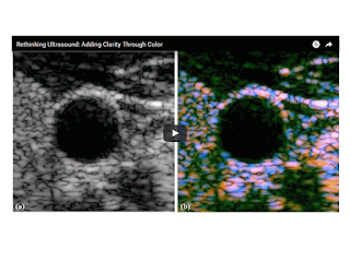
COLORING ULTRASOUND

 2016 Jun 21;61(12):L20-8. doi: 10.1088/0031-9155/61/12/L20. Epub 2016 May 25.

Scattering and reflection identification in H-scan images.


Abstract

Medical ultrasound imaging scanners typically display the envelope of the reflected signal on a log scale. The properties of this image and speckle patterns from collections of scatterers have a number of well-known disadvantages. One is the inability to differentiate between different scatterers that may have fundamentally different frequency-dependent scattering cross sections. This study proposes a framework for characterizing scattering behavior and visualizing the results as color coding of the B-scan image. The methodology matches a model of pulse-echo formation from typical situations to the mathematics of Gaussian weighted Hermite functions. The results show an ability to reveal some of the information otherwise hidden in the conventional envelope display, and can be generalized to more conventional bandlimited pulse functions. This new class of images is termed H-scan where 'H' stands for 'Hermite' or 'hue' to distinguish it from conventional B-scan format.

Inspired by a 19th-century set of mathematical functions, University of Rochester researcher Kevin Parker has devised a way to incorporate new color identifications in ultrasound medical images, making it easier to differentiate fine details that currently appear as indistinguishable objects in shades of gray.
The new imaging format would be especially valuable in helping physicians interpret ultrasound images of soft tissue, including muscle, glands, and organs such as the liver.
“This has been a great goal” of ultrasound research since the 1970s, said Parker, the William F. May Professor of Engineering. The technology, described in a paper in Physics in Medicine & Biology, provides more detailed soft tissue images than other attempts, based on quantitative backscattered imaging, Parker said.
Ultrasound uses pulses of high-frequency sound waves that bounce back echoes when they strike cells or arteries. These echoes are also known as scattered waves. The bouncing back gives the ultrasound image its features. With few exceptions – such as Doppler-enhanced ultrasound imaging of blood flow – those features consist of varying shades of black, white and gray reflecting different densities.
“If you look at an ultrasound image of the liver, there are so many things in there – veins, arteries, biliary ducts, liver cells, perhaps some scar tissue – and they’re all just displayed as black and white blobs,” Parker said. “If there’s a large artery, it’s easy to see the wall and the blood inside. But at the finer levels of detail, it is often impossible to tell if you’re looking at a smaller artery or 10 little cells.”
A set of mathematical functions — devised in 1890 by the great mathematician Charles Hermite of France and rarely used in engineering — provided Parker with a way to approach this problem. He came across the functions while perusing a handbook of transforms and applications, and immediately recognized that Hermite’s functions closely approximated ultrasound pulses.
“I realized if we used these, it would make our analyses of ultrasound scattering easier,” Parker said. “So now, instead of ultrasound images showing all of these tissue structures as black and white objects, we can now classify them mathematically (by their size) and assign unique colors to unique types of scatterers.”

Thứ Ba, 21 tháng 2, 2017

ARFI ĐÁNH GIÁ ĐỘ CỨNG CỦA GAN Ở BỆNH NHÂN TIỂU ĐƯỜNG TYPE 2

ARFI ĐÁNH GIÁ ĐỘ CỨNG CỦA GAN Ở BỆNH NHÂN TIỂU ĐƯỜNG TYPE 2  
NGUYỄN THỊ HỒNG ANH - NGUYỄN THIỆN HÙNG - PHAN THANH HẢI
TRUNG TÂM Y KHOA MEDIC HÒA HẢO
THÀNH PHỐ HỒ CHÍ MINH
ABSTRACT:

Using ARFI SIEMENS S2000 for measuring liver stiffness to grade the degree of liver fibrosis regardless liver steatosis. 80 adult type 2 diabetic patients were performed systematically to identify those with significant liver fibrosis. A significant liver stiffness increase was found in more than 40% of DM patients.
ARFI technique is comparable with TE in liver stiffness assessment.

ĐẶT VẤN ĐỀ
Dùng siêu âm đàn hồi ARFI SIEMENS S2000 đánh giá độ cứng gan (liver stiffness)  bệnh nhân tiểu đường, từ đó lượng giá mức độ xơ hóa của gan (fibrosis). Đánh giá độ thấm mỡ của gan (liver steatosis) bằng siêu âm B-mode.

ĐỐI TƯỢNG-PHƯƠNG PHÁP
ž  Đối tượng: bệnh nhân tiểu đường trên 18 tuổi và bình giáp, không đang mang thai hay cho con bú, không bị nhiễm HBV, HCV, không bị viêm gan tự miễn hay viêm gan do thuốc, không uống rượu bia quá 20g/ngày.
ž  Phương pháp:
·         Bệnh nhân được chẩn đoán tiểu đường và thỏa các tiêu chuẩn chọn bệnh.
·         Các xét nghiệm bao gồm đường huyết lúc đói, HbA1C, bilan lipid máu (LDL-Cholesterol, Triglyceride), creatinin, AST, ALT, GGT, FT4, TSH, HBsAg, AntiHBs, AntiHCV, Công thức máu. Xét nghiêm viêm gan tự miễn được thực hiện nếu men gan cao mà không nhiễm viêm gan siêu vi (LKM-1, AMA-M2, ANA).
·         Dùng siêu âm B-mode phát hiện và phân loại gan thấm mỡ S1, S2, S3  và không thấm mỡ S0.
·         ARFI gan được thực hiện sau siêu âm B-mode.
·         Thiết kế nghiên cứu cắt ngang, mô tả.

BẢNG PHÂN LOẠI GAN THẤM MỠ (B-MODE)


KẾT QUẢ
ž  Gồm 80 trường hợp bệnh nhân tiểu đường type 2 từ 28-79 tuổi, trong đó có 27 nam. Từ tháng 11-2016 đến tháng 02-2017.
ž  Được tổng hợp trong bảng kê dưới đây.
Số ca
S0
S1
S2
S3
Tổng
F0
5
6
6
0
17
F1
4
18
6
0
28
F2
2
3
6
1
12
F3
2
4
4
1
11
F4
0
5
4
3
12
Tổng
13
36
26
5
80

BÀN LUẬN
  Kỹ thuật không bị hạn chế khi bệnh nhân bị béo phì, báng bụng, khoang liên sườn hẹp.
  Xơ hóa gan đáng kể (F2-F3): 28,75% (23 ca) ở bệnh nhân có hoặc không có gan thấm mỡ.
  Xơ hóa gan nặng (F4):  15% (12 ca) đều ở nhóm bệnh nhân có gan thấm mỡ .
  100% gan thấm mỡ nặng (S3: 5ca) có xơ hóa gan nặng/đáng kể .
  5% (4 ca) có xơ hóa gan đáng kể (F2-F3) mà không có gan thấm mỡ : ghi nhận ở bệnh nhân có thời gian mắc bệnh lâu ngày (7-15 năm).

KẾT LUẬN
  Kỹ thuật ARFI là phương tiện nhanh có giá trị giúp đánh giá độ cứng gan ở bệnh nhân tiểu đường.
  Độ tin cậy cao trong xác định bệnh nhân không có hay có xơ hóa gan mức độ trung bình (F0 đến F2) với bệnh nhân xơ hóa gan nặng (F3-F4).

TÀI LIỆU THAM KHẢO CHÍNH


1. Liver Stiffness Evaluation by Transient Elastography in Type 2 Diabetes Mellitus Patients with Ultrasound-proven Steatosis:Sporea I, Mare R, Lupușoru R, Sima A, Sirli R, Popescu A, Timar R.: J Gastrointestin Liver Dis, 2016, Vol. 25, No 2: 167-174.
2. Liver stiffness in nonalcoholic fatty liver disease: A comparison of supersonic shear imaging, FibroScan, and ARFI with liver biopsy, Christophe Cassinotto,Jérome Boursier,Victor de Lédinghen,Jérome Lebigot, Bruno Lapuyade,Paul Cales,Jean-Baptiste Hiriart,Sophie Michalak,Brigitte Le Bail,Victoire Cartier,Amaury Mouries,Frédéric Oberti,Isabelle Fouchard-Hubert,Julien Vergniol,Christophe Aubé. Hepatology, Vol 63, June 2016.
3. Principles and clinical application of ultrasound elastography for diffuse liver disease: Woo Kyoung Jeong, Hyo K. Lim, Hyoung-Ki Lee, Jae Moon Jo, Yongsoo Kim. Ultrasonography 33(3), July 2014.