Tổng số lượt xem trang

Thứ Bảy, 13 tháng 7, 2013

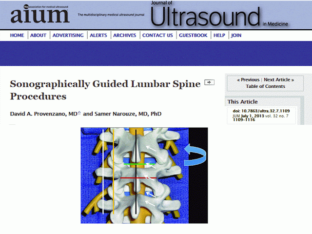
Lumbosacral Regional Anesthesia: Sonographic Guidance


CONCLUSION


     Sonographic guidance for lumbosacral regional anesthesia and chronic pain management procedures is rapidly evolving. An in-depth understanding of anatomy is important to enable correct identification of sonographically visualized structures. When compared to palpation-guided techniques, sonographically assisted procedures have been shown to improve clinical outcomes. When used for chronic pain management lumbar spine procedures, sonography has certain visualization advantages and limitations compared to fluoroscopy. Great strides have been made in the advancement of sonography as a primary visualization technique for specific lumbosacral spine procedures. Further advancements in sonographically guided scanning techniques and equipment development are needed to overcome some of the current sonographic visualization limitations for lumbosacral procedures. In addition, future studies are needed to evaluate the safety and efficacy of sonographically guided techniques for lumbosacral procedures.
Thứ Ba, 9 tháng 7, 2013

NHÂN CA Pseudoaneurysms (False Aneurysms) TẠI MEDIC

A pseudoaneurysm, or false aneurysm, is a collection of blood that results from a leaking hole in an artery. This life-threatening condition is described below.

Posted by | Last updated: Mar 25, 2013
 
A pseudoaneurysm (false aneurysm) results from a leakage of blood from an artery after trauma or after "dehiscence," or separation, of a surgical anastomosis. The causes are discussed in greater detail below. Blood leaks from the artery into the surrounding tissue to form the pseudoaneurysm. Since blood from the artery continues to fill the pseudoaneurysm, it is under relatively high pressure and can expand and compress the surrounding structures (nerves, veins, etc.).
This article is part of a three-part series. The causes, differential diagnosis, and diagnosis of pseudoaneurysms is described below. In Part 2, the Treatment of Pseudoaneurysms is discussed. Finally, Part 3 provides a Summary of Pseudoaneurysms and a listing of references.
 
Causes of Pseudoaneurysms
  • Penetrating trauma: A gunshot wound, stabbing, or bomb blast can disrupt the arterial wall and allow blood to leak into the surrounding tissue, forming a pseudoaneurysm.
  • Blunt trauma: A kick to the shin or even a bad ankle sprain can damage the wall of an artery enough to create a pseudoaneurysm.
  • Endovascular procedure: During certain medical (endovascular) procedures, doctors will introduce thin wires and instruments into the body, usually through the femoral blood vessels in the groin. One example of such a procedure is "arteriography," in which doctors insert a thin instrument through the femoral artery into the aorta in the abdomen. They then inject a contrast dye, which flows into the arteries in the legs. They then take images with an X-ray machine (fluoroscopy) to observe how the contrast and blood flow through the arteries in the legs, looking for any stenoses (narrowings) or other damages. Since this procedure involves injection into the high-pressure arteries, this minor damage to the arterial wall can allow leakage of blood and formation of a pseudoaneurysm.
Differential Diagnosis of Pseudoaneurysms
When deciding if a patient has a pseudoaneurysm, other diagnoses to consider are the following:
  • Hematoma
  • Abscess
  • Arteriovenous fistula (AVF)
  • Lymphadenopathy: Enlargement of lymph nodes that occurs with infections, lymphoma, and other cancers.
  • Lymphocele
  • Deep venous thrombosis: Blood clots in the leg veins.
  • Compartment syndrome
How to Diagnosis a Pseudoaneurysm (False Aneurysm)
A patient with a pseudoaneurysm will typically have had some type of traumatic event that could have damaged the patient's blood vessels. This could be a recent blunt or penetrating trauma, or an endovascular procedure (as described above). A pseudoaneurysm occurs when blood leaks from the artery days to even years after this traumatic event. The patient will complain of a mass near the site of the trauma that is pulsating, painful, and warm. It may be possible to feel the vibration or movement of blood within the mass (called a "thrill"). If the pseudoaneurysm is large enough to compress a nearby nerve, the patient may experience numbness or tingling (called "paresthesias") or pain.
Imaging Techniques:
  • Doppler ultrasound: Can detect the size and shape of the mass (pseudoaneurysm) and determine if there is bloodflow into it from a nearby artery.
  • Arteriography, CT angiography, and MR angiography: These three imaging techniques are very good at showing the anatomy of the arterial system. A dye (contrast agent) is injected into the bloodstream proximal to (before) the area of the pseudoaneurysm. Then an X-ray, CT scan, or MRI is obtained as the contrast dye flows through the arteries. As seen in the images below, contrast flowing into a pseudoaneurysm can easily be seen with these images.
Other Diagnostic Techniques:
  • Needle aspirate: If the doctor is unsure if a warm, painful mass is a hematoma, pseudoaneurysm, abscess (infection), cyst, or enlarged lymph node, the doctor can insert a needle into the mass and attempt to withdraw fluid. If the needle withdraws blood, then the mass could be a hematoma or a pseudoaneurysm.
The Treatment of Pseudoaneurysms is discussed next, followed by a summary of pseudoaneurysms.
References
  1. Aiyer S et al. Pseudoaneurysm of the posterior tibial artery following a closed fracture of the calcaneus. A case report. J Bone Joint Surg 2005. 87(10):2308-12.
  2. Aydin A, Lee CC, Schultz E, and Ackerman J. Traumatic inferior gluteal artery pseudoaneurysm: Case report and review of the literature. Am J Emer Med 2007. 25:488.e1-e3.
  3. Baynosa RC et al. Iatrogenic pseudoaneurysm following reverse radial forearm free tissue transfer. J Reconstructive Microsurgery 2007. 23(6):335-8.
  4. Corso R et al. Large iatrogenic pseudoaneurysm of the posterior tibial artery treated with sonographically guided thrombin injection. Am J Radiology 2003. 180:1479-80.
  5. Wikipedia: Pseudoaneurysm
From Suite 101