Tổng số lượt xem trang

Thứ Bảy, 6 tháng 7, 2024

HISTOTRIPSY based on ULTRASOUND


Abstract

Histotripsy is the first noninvasive, non-ionizing, and non-thermal ablation technology guided by real-time imaging. Using focused ultrasound delivered from outside the body, histotripsy mechanically destroys tissue through cavitation, rendering the target into acellular debris. The material in the histotripsy ablation zone is absorbed by the body within 1–2 months, leaving a minimal remnant scar. Histotripsy has also been shown to stimulate an immune response and induce abscopal effects in animal models, which may have positive implications for future cancer treatment. Histotripsy has been investigated for a wide range of applications in preclinical studies, including the treatment of cancer, neurological diseases, and cardiovascular diseases. Three human clinical trials have been undertaken using histotripsy for the treatment of benign prostatic hyperplasia, liver cancer, and calcified valve stenosis. This review provides a comprehensive overview of histotripsy covering the origin, mechanism, bioeffects, parameters, instruments, and the latest results on preclinical and human studies.

Keywords: High intensity ultrasound, ultrasound, physics, imaging, immunotherapy
Liver Histotripsy

The University of Chicago Medicine was the first health system in Illinois to treat patients with histotripsy, a new noninvasive technology that uses ultrasound energy to precisely destroy liver tumors.

UChicago Medicine has been involved for years in bringing histotripsy to patients, having treated patients in the US arm of the #HOPE4LIVER clinical trial. Histotripsy technology received FDA approval in October 2023, and UChicago Medicine physicians expect to begin clinical implementation in early 2024.

Safely Destroying Tumors With Targeted Sound Waves

Histotripsy technology uses high-intensity sound waves to selectively destroy cancer tissue in the liver. Unlike traditional methods such as surgery, radiation, or ablation, the procedure does not require any incisions, radiation, needles, or invasive procedures.

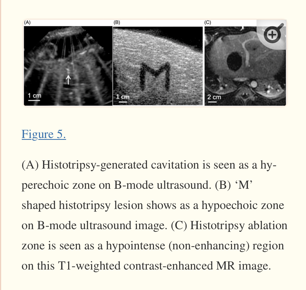
Before histotripsy treatment, doctors use ultrasound imaging – like what is used to see babies in the womb – to locate the tumor, determine the size of the area that needs to be treated, and target and monitor the treatment area during the procedure. After doctors program the histotripsy robot with the tumor’s coordinates, the machine precisely generates focused sound waves that create a "bubble cloud" inside the targeted area, which disrupts and destroys only the cancerous tissue.

“One of the best things about how this technology works is that it’s protective of things we don’t want to damage,” said Osman Ahmed, MD, associate professor of radiology at UChicago Medicine. “A histotripsy procedure can damage or kill a tumor while leaving important structures like blood vessels and bile ducts intact.”

The treatment is so simple and noninvasive that Ahmed said patients can even wear a t-shirt and jeans rather than a surgical gown.

“The actual procedure can take 5 to 30 minutes depending on the size of the tumor,” he said. “Then we move the robot away, and the patient wakes up. They can even go home the same day unless they have pain.”

Thứ Bảy, 29 tháng 6, 2024

LN-RADS - Lymph Nodes Reporting and Data System


Lymph nodes are crucial in the diagnosis and treatment of cancer. The LN-RADS system significantly improves the quality of lymph node assessment and, thanks to the support of artificial intelligence, it achieves an even higher level of consistency between the assessment and histopathological findings. AI solutions support both radiologists and oncologists in their daily work, and they can be an effective tool for education in this area.

LN-RADS US criteria recommendation ver.1.0

No enlargement (recommended max SAD up to 6-7mm), oval shape (L/S-ratio > 2), regular cortex, max. thickness ≤3mm, cortex echogenicity similar or higher to the background fatty tissue, smooth margins, no other changes in architecture (no calcifications, no fluid collections, no necrosis, no FCT), no pathological peripheral or chaotic vascularization.

LN-RADS 2 steatotic LN


LNs enlarged in one or both axes, cortex regular, max thickness ≤3mm, hyperechoic hilum (steatotic) with no size limits, no other changes in architecture (no calcifications, no fluid collections, no necrosis, no FCT, no pathological peripheral or chaotic vascularization).

LN-RADS 3 reactive LN


Probably due to the inflammatory process or vaccination. Dominant feature: regular, thickened cortex >3mm, enlargement in one or two axes, preserved oval shape (L/S-ratio > 2), preserved medulla, no other changes in architecture (no calcifications, no fluid collections, no necrosis, no FCT), cortical echogenicity similar to or moderately lower than the background fatty tissue, well-defined margins, no pathological peripheral or chaotic vascularization, no oncological or hematological history, no laboratory oncological abnormalities.

LN-RADS 4 suspicious LN


4a low, 4b high probability of malignancy. This group is dedicated to LNs that morphologically do not match groups 1, 2, 3, 5 or have additional radiological or clinical factors increasing the probability of malignancy in LNs categorized as LN-RADS 3, i.e. high or increasing laboratory markers (i.e. PSA for inguinal LNs), active neoplasm in the region (i.e. breast cancer for axillary LNs), other metastatic or systemic LNs in the region, clinical symptoms suggesting oncological or systemic hematological disease. The primary principle for categorizing LNs as group 4 is that “better check than miss”.

LN-RADS 4a 

Low suspicion of malignancy – the size may be normal in SAD and LAD, cortex with thickening ≤4mm, moderate irregularity, especially local. As a general assumption, all LN 4a should be verified by biopsy or PET, and if it is not possible, they should be treated as suspicious and malignant.    

LN-RADS 4b 

High suspicion of malignancy – the size may be normal in SAD and LAD, cortex  thickening >4mm and irregularity, especially FCT, or no hilum, shape more round than oval (L/S-ratio≤2), hypoechogenicity to the background fatty tissue, especially “black hole sign”, microcalcifications, fluid collections, necrosis,  abnormal peripheral or chaotic vascularization; ill-defined, blurred margin.

LN-RADS 5 malignant LN


Enlargement in SAD and one or more features of malignancy: lack of hilum, hypoechogenicity to the background fatty tissue or “black hole sign”, evident cortex irregularity /FCT”, shape more round than oval (L/S-ratio≤2), microcalcifications, fluid collections, necrosis, abnormal peripheral or chaotic vascularization; ill-defined, blurred borders or signs of extracapsular infiltration.

If evident radiological and clinical features of malignancy are present, enlargement is not necessary for LN-RADS 5 classification.